Douglas F. Milam M.D.
Urologic Surgery
- Home
- About Us
- Surgical Procedures

- Evaluation of Voiding Dysfunction
- Patient Information
- IPSS-AUA Symptom Score
- Sexual Function Questionnaire
- Deactivate a Urinary Sphincter
- VPEC
- Overnight Surgical Stay
- Post-operative Instructions Urethral Stricture Repair
- Post-operative Instructions Penile Implant
- Post-operative Instructions Artificial Urinary Sphincter
- Post-operative Instructions Placement of Male Sling
- Post-operative Instructions Penile Curvature Repair
- Post-procedure Instructions Vasectomy
- Mechanism of Erection
- Statement on Urologic Pain
- Schedule a Consultation
- Directions
- Urologic Images
- Web References
Cystoscopy

This image shows the normal prostate. You can see that the image quality is excellent. The "bump" slightly out of focus in the foreground is the veru montanum. This is a landmark urologists use to identify the prostatic apex. Small blood vessels can be easily seen due to the magnification of the optical system. In reality these vessels are about the size of a point of a pin.
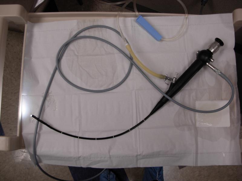
Cystoscopes are either flexible or rigid. We use flexible cystoscopes (shown below in the top photo) in the office practice under local anesthesia since they are more comfortable for the patient than rigid cystoscopes. Rigid cystoscopes (shown in the bottom photo) have better optics and larger working channels that allow laser fibers and other instruments to be passed within the sheath of the scope. Rigid cystoscopes are extremely useful for more complex procedures performed in the operating room.
Flexible cystoscope (Black) and the fiber optic light system. The small diameter and flexibility of the instrument allow for evaluation to be performed in the office.

Rigid cystoscopic equipment with working ports.